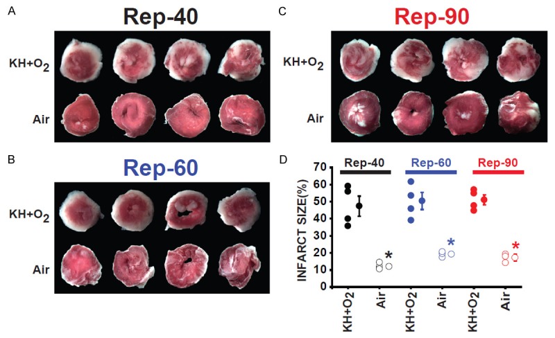
Figure 4.

Myocardial infarct size was much smaller in hearts suspended in air compared to hearts immersed in KH+O2 during the normothermic ischemic period at the end of the reperfusion. Images of four slices of different regions (four transverse slices parallel to the atrioventricular groove) from the same heart in Rep-40 (A), Rep-60 (B), and Rep-90 (C) groups of hearts immersed in KH compared with hearts suspended in air during the ischemic period and reperfused for the same duration. Hearts were stained with TTC, showing infarcted regions in white and viable healthy regions in red. (D) Bar graph showing the individual percentage of myocardial infarct size in Rep-40, Rep-60, and Rep-90 in both conditions of ischemic conservation. Infarct size did not change significantly after the different times of reperfusion between the three groups immersed in KH as well as the groups suspended in air during the ischemic insult. However, the myocardial infarct size was much smaller in the groups of hearts suspended in air when compared to hearts immersed in KH during the ischemic period. Values mean ± SEM.; *P<0.05 hearts immersed in KH+O2 versus hearts maintained in air during ischemia (n = 4-5/group).
