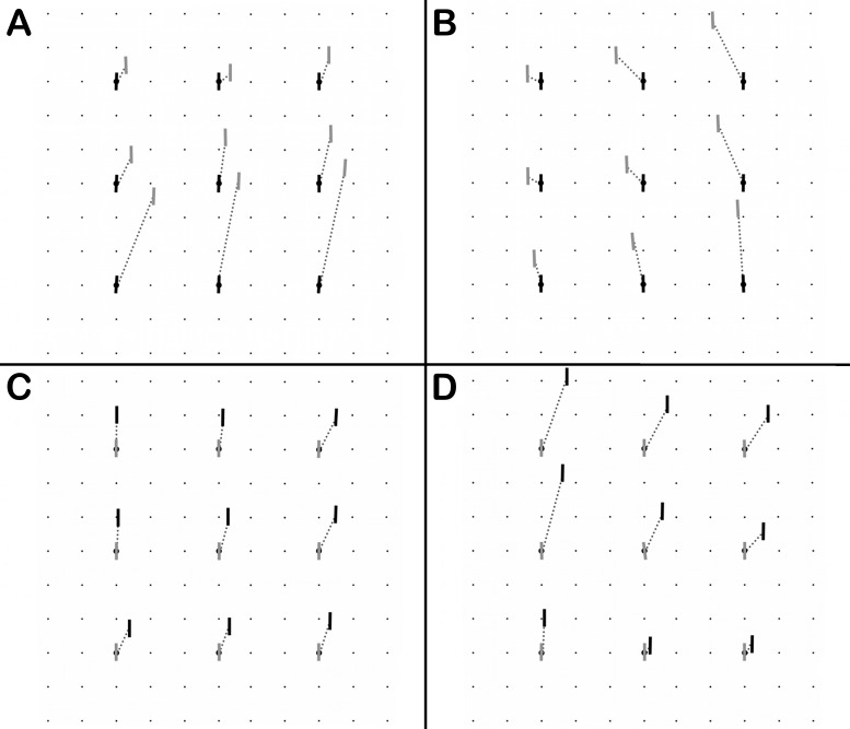
Figure 2.
Lancaster red-green plots from patients with “congenital SOP.” The black lines correspond to the projections of the subjective vertical meridians of the right eye onto the wall, and the gray lines correspond to the projections of the subjective vertical meridians of the left eye onto the wall. (A) Patient with vertical-rectus-muscle–mediated fusion, demonstrating an incomitant hyperdeviation that increases in down gaze (patient 1 shown). (B) Another patient with vertical-rectus-muscle–mediated vertical fusion, showing a left hyperdeviation that increases in adduction of the higher, left eye (also interpretable as a relatively comitant cyclovertical deviation, combining extorsion and elevation, of the left eye's approximately square eye movement pattern; patient 6 shown). (C) Patient with oblique-muscle–mediated fusion, showing general spread of comitance (patient 8 shown). (D) Patient with mixed (oblique/rectus muscle) mechanism of vertical fusional vergence, showing a right hyperdeviation that increases in upgaze and adduction of the higher, right eye (also interpretable as extorsion and elevation of the right eye's entire eye movement pattern; patient 10 shown).

