
Fig. 4. Phenotype alteration at 3 dpf in embryos treated with increasing concentrations of GSI A or GSI 18.
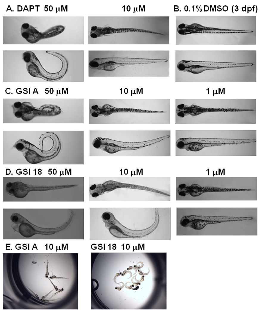
A. Embryos treated with 50 µM DAPT showed a strong phenotype, and much less effect was observed at lower concentration of 10 µM. B. DMSO treated embryos did not reveal any phenotype at 3 dpf. C. GSI A was used at 50 µM, 10 µM, and 1 µM. In contrast to embryos treated with 50 µM GSI A, 10 µM and 1µM of GSI A did not cause phenotype alteration. D. All embryos showed clear curved tails in 50 µM GSI 18. Embryos treated with 10 µM of GSI 18 still showed similar morphologic changes, and much less effect was observed at lower concentration of 1µM. E, All embryos treated with 10 µM GSI A showed normal phenotype similar to DMSO treated embryos. On the contrary, all embryos treated with 10 µM GSI 18 showed much stronger phenotypic alteration.