
Figure 3.
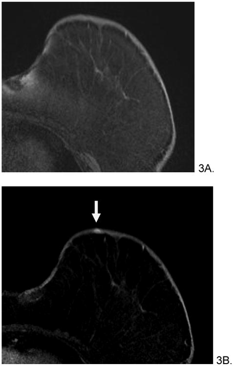
66 year old female with history of invasive ductal and lobular carcinoma of left breast, status post lumpectomy and radiation therapy 7 years prior who presented with left breast violaceous skin lesion. Skin punch biopsy revealed angiosarcoma. Pre-treatment breast MRI revealed T1 isointense (A), T2 heterogenous (not shown), focus of rapid enhancement (B, arrow). The patient underwent pre-surgical chemotherapy with taxol and gemcitabine before mastectomy. She is currently disease free.