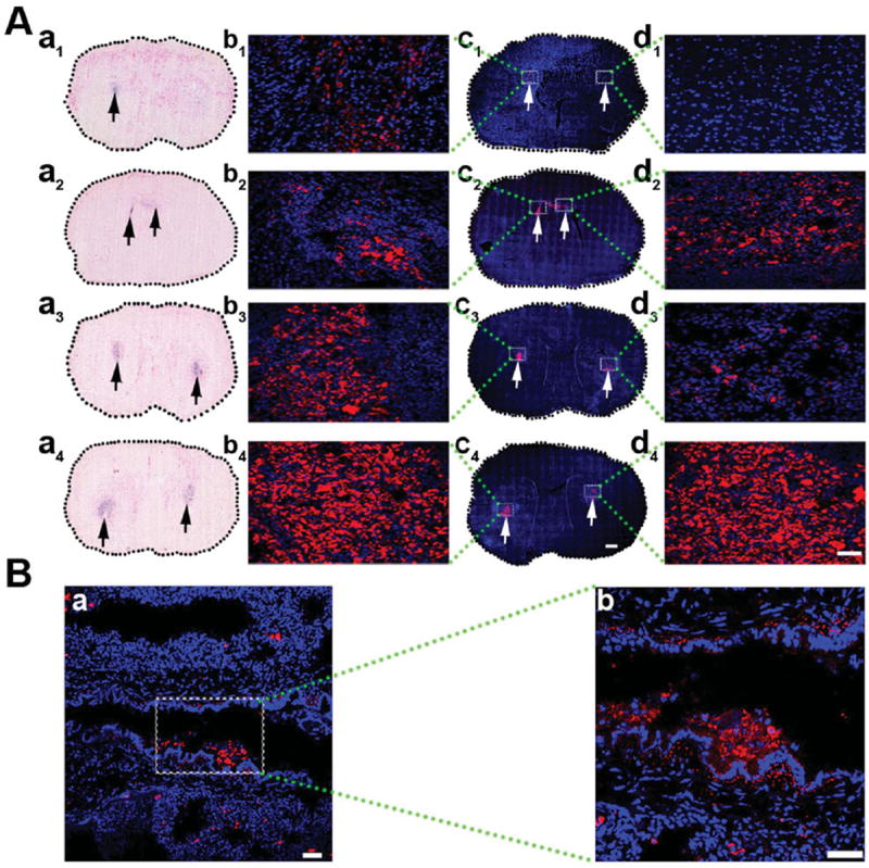
Figure 6.

Histological analysis of (125)I-fSiO4@SPIO-labeled MSCs in brain and lung. A) Prussian blue staining and confocal images of brain sections at day 1 (a1, c1), day 3 (a2, c2), day 7 (a3, c3), and day 14 (a4, c4) post IC transplantation of (125)I-fSiO4@SPIO-labeled MSCs. a1–a4) Prussian blue staining; c1–c4) confocal images. Scale bar = 500 μm. The left is the injection side and the right is the ischemic side. Arrows indicate the localization of labeled MSCs. (b1–b4) and (d1–d4) are magnified views of coronal sections corresponding to the white boxes in c1–c4. Scale bar = 50 μm. B) Confocal images of lung after 14 days of IV injection of (125)I-fSiO4@SPIO-labeled MSCs (a, red) in ischemic rats. Cell nuclei were counterstained with DAPI (blue). Scale bar = 50 μm. b) Magnified views of boxed area in a. Scale bar = 100 μm.
