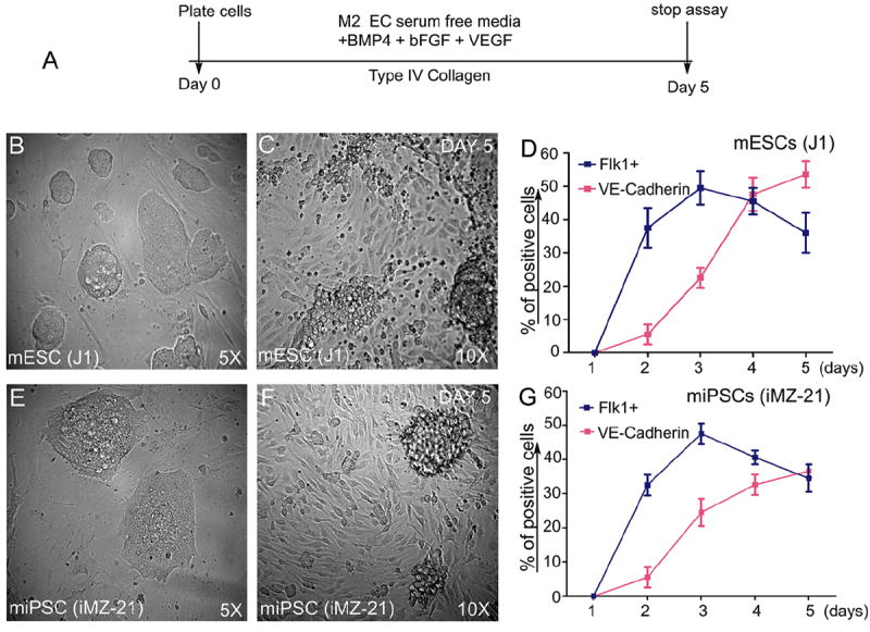
Figure 1. Induction of Flk1+VE-cadherin+ vascular EC progenies from iPS and ES cells.

Timeline of emergence of Flk1+VE-cadherin+ vascular ECs. (A). Undifferentiated mES (J1 line) or miPS (iMZ-21) cells were cultured for 5 d in IV Col-coated dishes in media containing BMP4, bFGF, and VEGF165 to induce generation of vascular EC progenies. (B&C). Representative phase contrast microscopy of mES cell-derived adherent vascular progenies after d 5 in culture at indicated magnifications (B&C). Representative phase contrast microscopy of miPS cell-derived vascular progenies after day 5 in culture at the indicated magnifications (E&F). FACS profile of the emergence of Flk1+VE-cadherin+ vascular progenies (D&G). All experiments were repeated >5 times. Data indicate the mean ± S.E.M. n=5. (Reprinted from Kohler EE et al., PloS One 2013 Dec 30;8(12):e85549).
