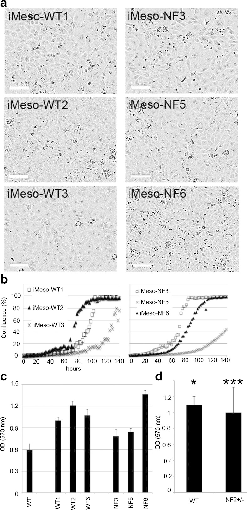
Figure 2.
Immortalized mesothelial cell morphology and cell growth in DMEM + 10% FBS. (a) Brightfield images were taken with the Incucyte Live Cell Imaging system (FLR 10×) and show immortalized mesothelial cell clones derived from WT and Nf2+/− mice. The typical “cobblestone-like” morphology is conserved; all clones show a higher proliferation rate than their nonimmortalized primary counterparts (scale bar: 100 μm). (b) Representative Incucyte growth curves of WT and Nf2+/− clones. (c) MTT signals obtained at 144 h postseeding using all cells and cell lines as in Fig. 1 and 2a. (d) MTT data grouped for three clones each of the genotypes WT and Nf2+/− in comparison to the primary mesothelial cells (*p < 0.05; ***p < 0.0005).

