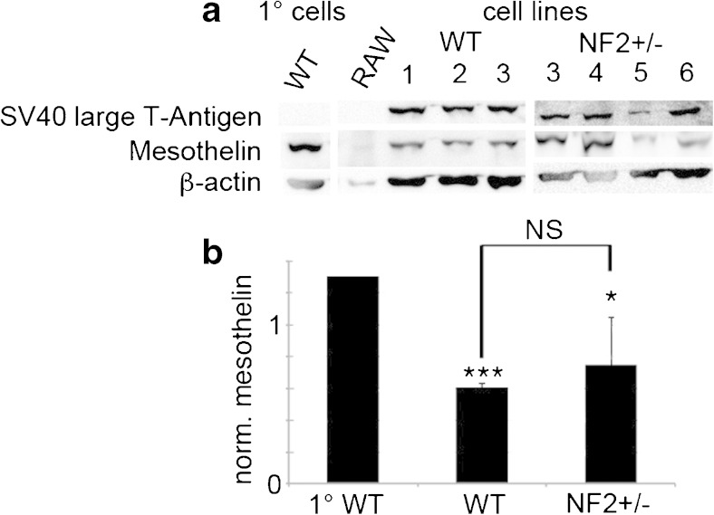
Figure 3.
Western blot for SV40 large T antigen and mesothelin in WT and Nf2+/− cells in comparison of the primary immortalized clones from WT and Nf2+/− mesothelial cells. (a) Western blot signals from a representative experiment. The Western blot signals for β-actin were used for the normalization of the mesothelin expression levels. (b) Quantitative analysis of a representative Western blot (n = 3 samples per genotype, p < 0.05 grouped primary cells vs. grouped immortalized cell lines).

