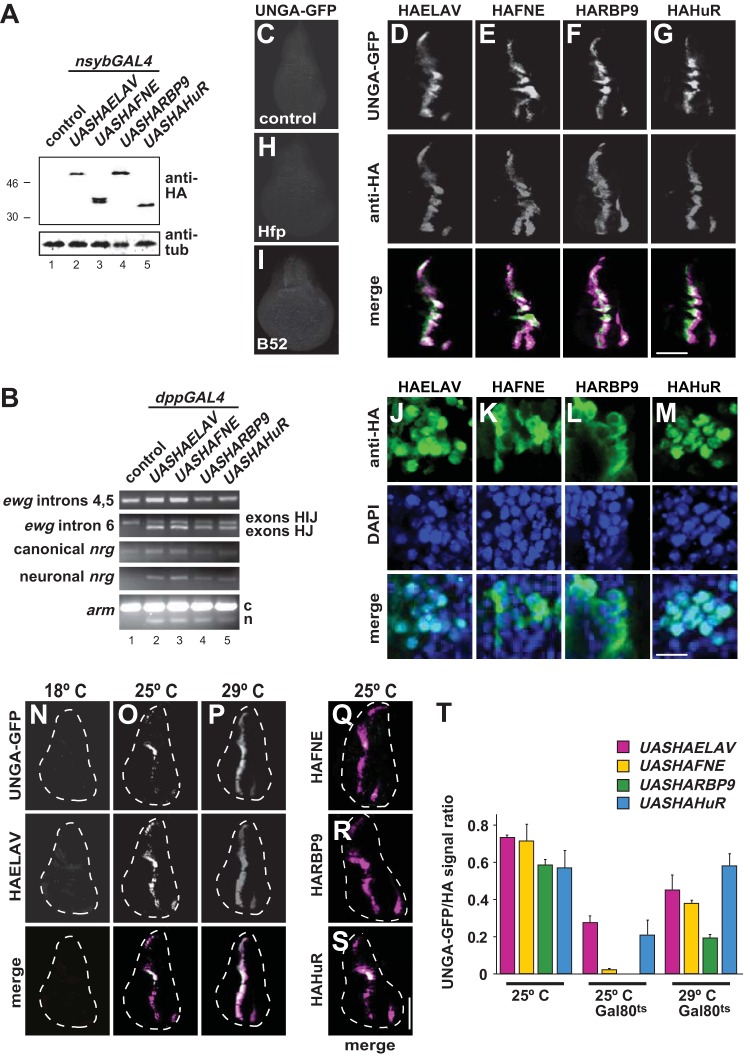
FIG 4.
Elevated levels of FNE, RBP9, and HuR can regulate alternative splicing of ELAV targets. (A) Expression of HA-tagged ELAV (e.g., HAELAV), FNE, RBP9, and HuR from UAS-containing transgenes in adults with nsyb-GAL4 by Western blotting detection with anti-ELAV antibodies. (B) Neuronal alternative splicing of ELAV targets ewg intron 6 from exon H to J, nrg and arm induced by expression of HA-tagged ELAV, FNE, RBP9, and HuR from UAS-containing transgenes in wing discs with dpp-GAL4 as assessed by RT-PCR. c, canonical; n, neuronal. (C to I) Neuronal alternative splicing of the nrg GFP reporter UNGA upon expression of HA-tagged ELAV, FNE, RBP9, and HuR from UAS-containing transgenes in wing discs with dpp-GAL4. Staining with anti-GFP and anti-HA is as indicated on the left. Due to temporally regulated expression of dpp-GAL4 and because expression of ELAV proteins precedes GFP expression, signals of ELAV proteins and GFP do not entirely overlap. Note that the distantly related poly(U) binding protein Hfp (H) and the SR protein B52 (I) do not induce UNGA splicing. Scale bar, 150 μm. (J to M) Cellular localization of HA-tagged ELAV, FNE, RBP9, and HuR from UAS-containing transgenes in wing discs with dpp-GAL4. Staining with anti-HA and DAPI is as indicated on the left. Scale bar, 10 μm. (N to S) Neuronal alternative splicing of the nrg GFP reporter UNGA upon expression of HA-tagged ELAV, FNE, RBP9, and HuR from UAS-containing transgenes in wing discs with dpp-GAL4 in the presence of temperature-sensitive inhibitor of GAL4, GAL80ts, expressed from a UAS transgene at 18°C, 25°C, and 29°C. Staining in panels N to P with anti-GFP and anti-HA is as indicated on the left. Merged images are shown in the bottom row of panels N to P and in panels Q to S. Scale bar, 150 μm. (T) Quantification of UNGA-splicing shown as means with the standard error from five wing discs.

