Figure 6.
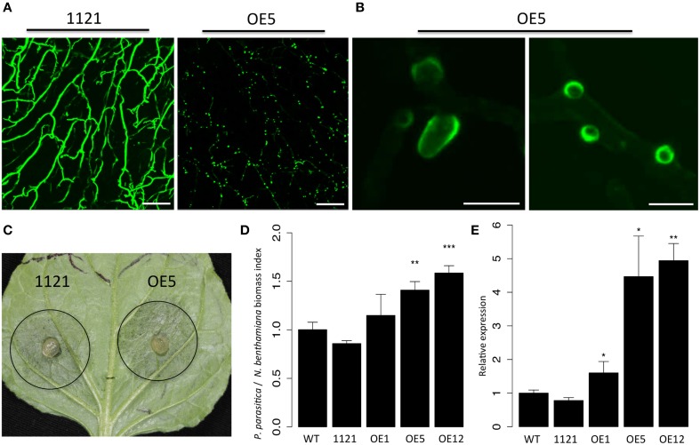
PpPDI1 is associated with haustoria-like structures of P. parasitica during colonization of N. benthamiana. (A) Increased number of haustoria-like structures produced by P. parasitica transformant OE5 expressing PpPDI1-EGFP on infected N. benthamiana leaf tissue compared with the control transformant 1121 (bar, 100 μm). Confocal laser scanning microscopy was used to define hyphal cytoplasm and haustoria-like structures. (B) The PpPDI1-EGFP protein is highly enriched at periphery of the haustoria-like structures of P. parasitica during plant infection (bar, 10 μm). Confocal laser scanning microscopy was used to define the haustoria-like structures. (C) Leaves of 6-week-old N. benthamiana infected with P. parasitica transformant OE5 and the control transformant 1121. The leaves were inoculated with agar plugs containing P. parasitica mycelia. The photo was taken at 60 hpi. (D) Real-time PCR assay to determine P. parasitica biomass in infected plant tissues following inoculation with P. parasitica. Total genomic DNA from P. parasitica infected tissues was isolated at 60 hpi. Real-time PCR employed primers specific for the N. benthamiana and P. parasitica constitutively genes. Bars represent standard errors from three biological replicates. (E) Real-time RT-PCR assay is used to quantitate PpPDI1 expression in P. parasitica wild-type recipient strain Pp016, the control transformant 1121, and three transformants expressing PpPDI1-EGFP (OE1, OE5, and OE12) during infection of N. benthamiana. The infection tissues were sampled 60 hpi. The real-time RT-PCR experiments were repeated three times with independent RNA isolations. Bars represent standard errors from three biological replicates. P. parasitica WS041 was used as a reference gene for quantification and normalization of PpPDI1 expression. (*P < 0.05; **P < 0.01; ***P < 0.001).

