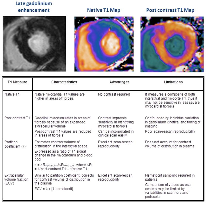
FIGURE 5.
To date, there are four different myocardial T1 mapping techniques used to assess diffuse interstitial fibrosis. Each technique has its own unique merits and limitations. In aortic stenosis, extracellular volume fraction appears to be the most promising technique in assessing diffuse fibrosis. Extracellular volume fraction demonstrates excellent scan-rescan reproducibility, which is necessary when assessing change related to treatment response and disease progression.

