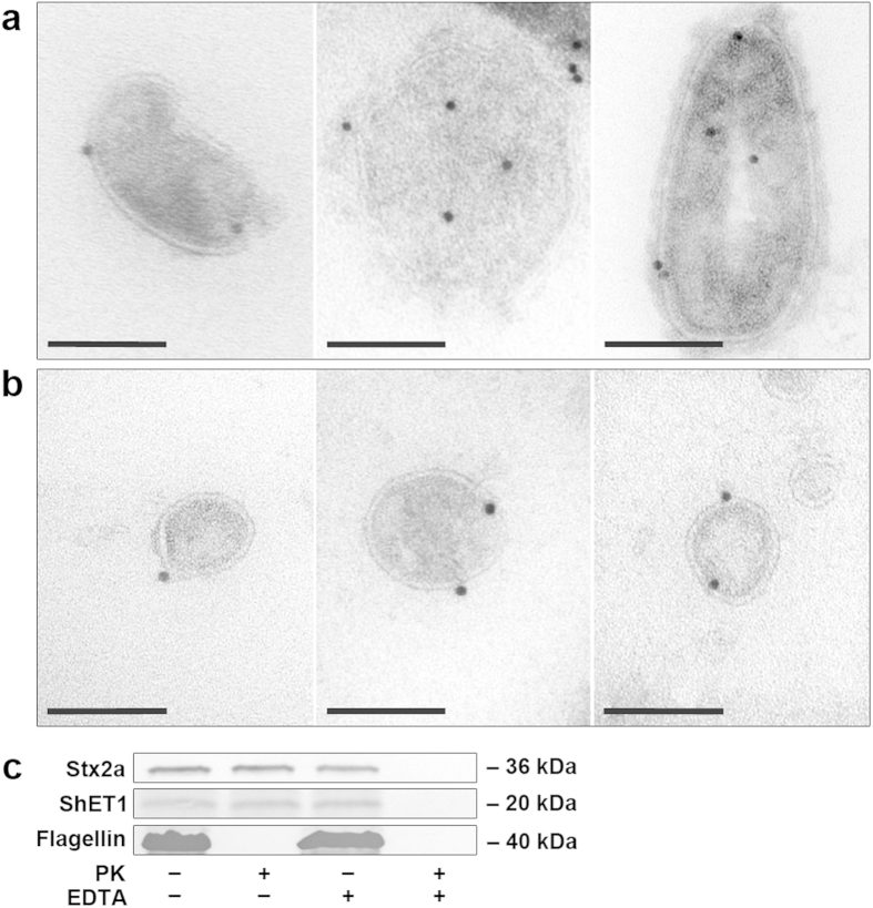
Figure 3. Localisation of Stx2a, ShET1 and H4 flagellin within OMVs.
(a,b) Immunogold staining of ultrathin frozen sections of purified OMVs from strain LB226692 with (a) anti-Stx2a and (b) anti-H4 antibody and Protein A Gold (10 nm). The images were acquired with a FEI-Tecnai 12 electron microscope. Bars are 100 nm. (c) Immunoblot analyses of proteinase K (PK)-untreated (PK−) and PK-treated (PK+) OMVs LB226692 either intact (EDTA−) or lysed with 0.1 M EDTA (EDTA+) with anti-Stx2a, anti-ShET1, and anti-H4 flagellin antibodies. Signals were visualised with Chemi Doc XRS imager. Sizes of immunoreactive bands are shown on the right side. Crops of representative immunoblots are shown. Full immunoblots are shown in Supplementary Fig. S8.

