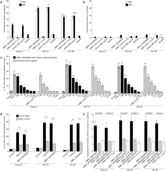
Figure 6. OMV-associated Stx2a causes apoptosis of human IECs via caspase-9 and caspase-3 activation.
(a,b) Caco-2, HCT-8, and HT-29 cells were incubated with OMVs LB226692 (containing 580 ng/ml of Stx2a) or purified Stx2a (580 ng/ml; control) or OMVs C227-11Φcu or OMV buffer for 24 h and 48 h or remained untreated; cells were analysed for apoptosis (a) and necrosis (b) by Cell Death Detection ELISA. Enrichment factors were calculated by dividing OD405 absorbance values of sample-treated cells with those of untreated cells; **P < 0.01 compared to OMV buffer (one-way ANOVA). (c) Cells were incubated (48 h) with decreasing doses of OMVs LB226692 (containing the indicated amounts of Stx2a) or with the same doses of purified Stx2a, or with OMVs C227-11Φcu (Stx2a-negative) or 1 μM staurosporin (Stauro; positive control) or remained untreated (control). Cells with hypodiploid nuclei were quantified by flow cytometry (FACScalibur) after propidium iodide staining (red channel, 570 nm). Data from 10,000 nuclei were analysed by CellQuestTM Pro software. **P < 0.01, and *P < 0.05 compared to untreated cells (one-way ANOVA). (d) Cells were incubated (48 h) with OMVs LB226692 (containing 580 ng/ml of Stx2a) or 1 μM staurosporin (Stauro; positive control) without and after cell pre-treatment with pan-caspase inhibitor z-VAD-fmk. Apoptotic cells were quantified as in c; **P < 0.01, and *P < 0.05 for comparison between non-pretreated and z-VAD-pretreated cells (unpaired Student´s t test). (e) Cells were incubated (48 h) with OMVs LB226692 (containing 580 ng/ml of Stx2a) or OMVs C227-11Φcu (Stx2a-negative) or remained untreated. Caspase-9 and caspase-3 activities in cell lysates were determined using colorimetric substrates (LEHD-pNA and DEVD-pNA, respectively); the colour intensity, which is proportional to the level of caspase enzymatic activity, was measured spectrophotometrically at 405 nm (FLUOstar OPTIMA reader). The caspase activity in OMV-treated cells was expressed as a fold-increase of that in untreated control cells (set up as 1). Inhibitors of caspase-9 (z-LEHD-fmk) or caspase-3 (z-DEVD-fmk) were added to cells 30 min before OMVs; *P < 0.05 compared to untreated cells (one-way ANOVA). Data in all panels are shown as means ± standard deviations from three independent experiments.

