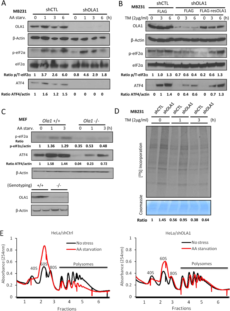
Figure 3. OLA1 regulates ISR signaling in cancer cells.
(A) The shCTL and shOLA1 cells of MB231 origin were AA-starved for the indicated time and analyzed by WB. (B) Cells were transfected with FLAG-only or FLAG-resOLA1 plasmids for 48 h, then treated with 2 μg/ml TM. (C) Ola1 (+/+) and (−/−) primary MEF cells were cultured in AA free medium for the indicated time. Genotyping of embryos used for MEF isolation was done by both PCR and IB (bottom). (D) Analysis of de novo protein synthesis in MDA-MB-231 shCTL and shOLA1 cells under ER stress. Cells were treated with TM for the indicated time and pulse-labeled with [35S]Met/Cys. Total proteins were separated on SDS-PAGE and autoradiographed. The [35S] incorporation (top) is normalized for loading of proteins indicated by Coomassie blue staining (bottom). (E) Polysome profiling of HeLa shCtrl and shOLA1 cells with or without 6 h AA starvation. Full scan images of immunoblots are presented in Figure S12.

