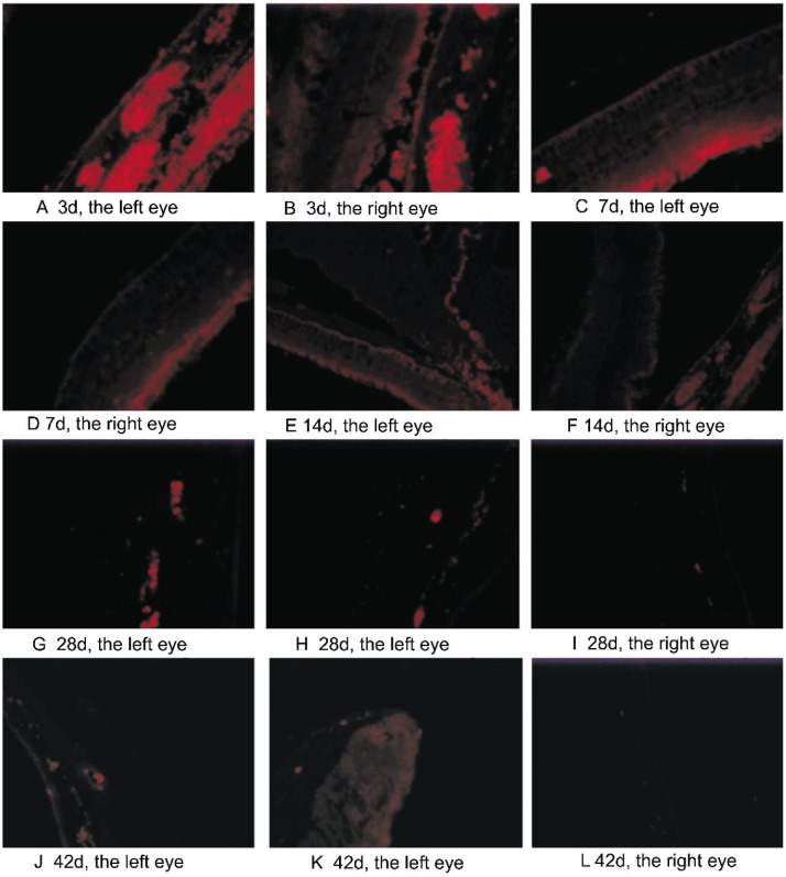
Figure 4. The distribution of bevacizumab in the eye tissues. The immunofluorescence staining can be observed in the retina, choroid, iris, ciliary body and anterior chamber angle, especially in the vascular tissues after intravitreal injection.
Strongest staining was seen at day 3 and day 7 (A-D) in both eyes. The intensity of staining in the left eyes was weakened to bright staining at day 14 and day 28 (E, G). Faint staining could also be detected at day 42 (J, K). Contrastively, in the right eyes, bright staining was seen at day 14 (F) which was weaker than the left eyes. Only very weak staining was detected at day 28 (I), and almost no staining can be seen at day 42 (L).

