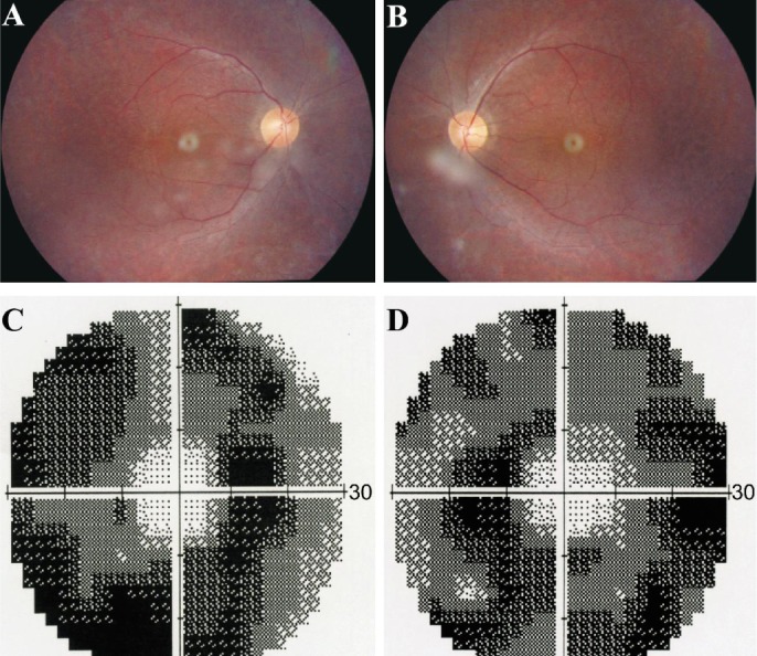
Figure 3. Bilateral fundus photography and visual field of the proband.

A, B: Bilateral fundus photography showing atrophy of the retinal pigment epithelium and attenuation of retinal arterioles, while the optic disc color were normal of the both eyes of the proband; C, D: Bilateral vision field got concentric narrowing, for the residual central visual field were 10 degrees.
