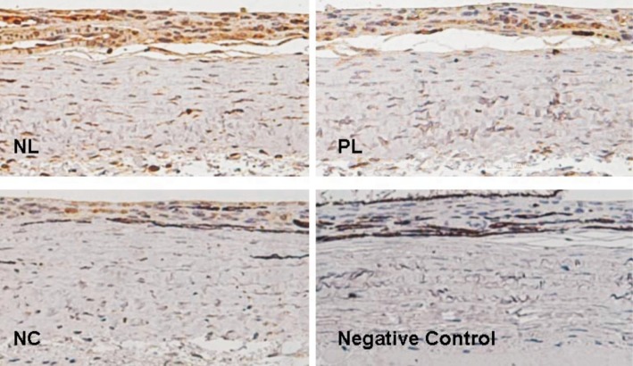
Figure 1. C5b-9 immunostaining in the posterior sclera.
Positive cells showed brown staining. Immunopositive staining of C5b-9 was increased in the posterior sclera of NL group eyes, compared to those of the PL and NC groups; no C5b-9 staining was observed when the primary antibody was omitted (negative control).

