
Fig. 1.
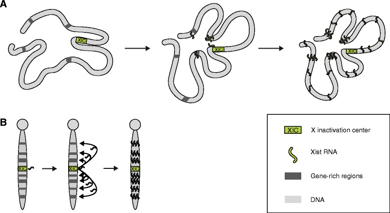
Models of the localization and spreading of Xist. a Three-dimensional spreading model of Xist localization. Xist might use close-proximity sites for its initial spreading (left and middle panels) before accumulating over the whole chromosome. At the final stages of spreading, Xist shows the highest enrichment at gene-rich regions (right panel). b Linear model of Xist spreading showing a classical representation of Xist decorating G-light bands on metaphase chromosomes