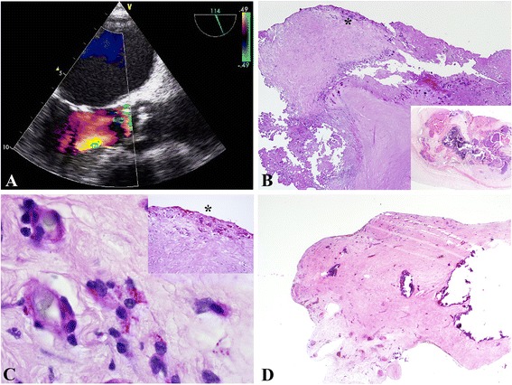
Fig. 1.

a–d: Whipple endocarditis and pericarditis in a 55-year old patient. a. TEE showing a severe degenerative aortic valve and aortic valve vegetations with aortic regurgitation III°. b. Severely degenerative and calcific aortic disease with chronic ulcerations (Pas, 2x, inset: hematoxillin&eosin, 2x). *labels the area visualized in 1C. c. Demonstration of Pas-reactive foamy macrophages, which contained T. whipplei particles. Note: the macrophages are seen in relation to capillaries (Pas, 100x, inset: Pas, 10x). d. Pericardium with extensive sclerosis and calcification (hematoxillin&eosin, 2x)
