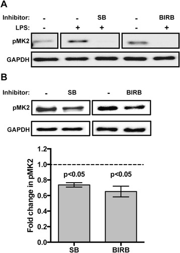
Fig. 3.

Inhibition of AsP38 MAPK reduced phosphorylation of MAPK-activated kinase 2 (MK2) in vitro and in vivo. a Representative western blot of phosphorylated MK2 (pMK2) from ASE cells pretreated with 10 μM SB203580 (SB), 10 μM BIRB796 (BIRB), or an equivalent volume of DMSO as a control for 2 h and then stimulated with 1 μg/ml LPS for 15 min. GAPDH provided an assessment of protein loading. b Representative western blot of phosphorylated midgut MK2 (pMK2) from A. stephensi fed a P. falciparum-infected blood meal supplemented with 10 μM SB203580 (SB), 10 μM BIRB796 (BIRB), or an equivalent volume of DMSO; tissues were dissected at 2 h post-feeding for analysis. Graph represents fold change ± SEMs of pMK2 protein levels normalized to untreated controls, n = 3. Pairwise comparisons of treatments versus controls at each time point were analysed by Student’s t-test, significant p-values are shown
