Figure 1.
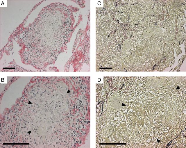
Lung extracellular matrix destruction and caseous necrosis colocalize in human pulmonary granulomas. Lung biopsy specimens from patients who were under investigation for lung carcinoma but received a final diagnosis of tuberculosis on the basis of histological findings were stained by Picrosirius red (A and B; collagen fibrils stain red), and elastin–van Gieson (C and D; elastin fibrils stain blue). Arrowheads designate areas of caseous necrosis. Collagen and elastin fibrils are absent in all regions of caseous necrosis. Images are representative of lung biopsy specimens from 5 patients with tuberculosis. Scale bars, 100 µm.

