Figure 2.
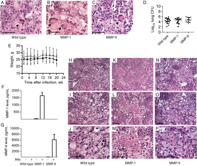
Mice expressing human matrix metalloproteinase 1 (MMP-1) develop regions of caseous necrosis in tuberculosis granulomas. Mice expressing human MMP-1 or MMP-9 and their wild-type littermates were infected with an Indo-Oceanic strain of Mycobacterium tuberculosis recently isolated from a patient with pulmonary tuberculosis. Mice were euthanized 22 weeks after infection. A–C, All mice strains developed multinucleate giant cells in regions of macrophage infiltration (arrowheads). D, No difference in mycobacterial growth was observed between mice strains. Horizontal lines denote mean values, with bars denoting standard deviations (SDs). E, Mouse weights did not differ between strains during the course of infection. Circles denote wild-type mice, squares denote MMP-1–expressing mice, and triangles denote MMP-9–expressing mice, with mean values and SDs denoted by lines and whiskers, respectively. F and G, Infection upregulated human MMP-1 and MMP-9 in lung homogenates of the respective transgenic mice. Mean values plus standard errors of the mean are shown. H–P, In the MMP-1–expressing mice, regions of tissue destruction developed (arrowheads), with amorphous central material typical of human caseous necrosis (K, L, and M), which was not observed in similar granulomas in wild-type mice (H, I, and J) or MMP-9–expressing mice (N, O, and P). The experiment was performed 3 times, with a minimum of 5 mice per group. Scale bars, 25 µm. Abbreviation: CFU, colony-forming units.

