
Figure 4.
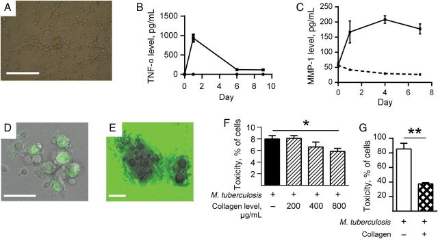
Collagen improves survival of Mycobacterium tuberculosis–infected cells in a 2-dimensional primary human cell culture system. Primary human peripheral blood mononuclear cells were infected with M. tuberculosis H37Rv in 24-well tissue culture plates and observed for 15 days. A, Cellular aggregates developed in M. tuberculosis–infected wells by day 4. B and C, M. tuberculosis infection increases secretion of tumor necrosis factor α (TNF-α) and matrix metalloproteinase 1 (MMP-1) in cell culture supernatants analyzed by Luminex array. D, Aggregates cause pericellular collagen destruction, analyzed by coculture with DQ-labeled collagen, which gains fluorescence when cleaved (E), or with fluorescent collagen, which loses fluorescence when degraded. F, Addition of collagen to M. tuberculosis–infected cells reduces cell death, as analyzed by the lactate dehydrogenase release assay. G, In a 3-dimensional model in which cells and M. tuberculosis are incorporated into an agar matrix with or without addition of collagen, incorporation of collagen with cells improves cellular survival after M. tuberculosis infection. Each experiment was performed a minimum of 2 times. Charts demonstrate the mean values + standard errors of the mean of a representative experiment performed in triplicate. Scale bars, 100 µm (A) and 25 µm (D and E). *P < .05, **P < .01.