Fig. 3.

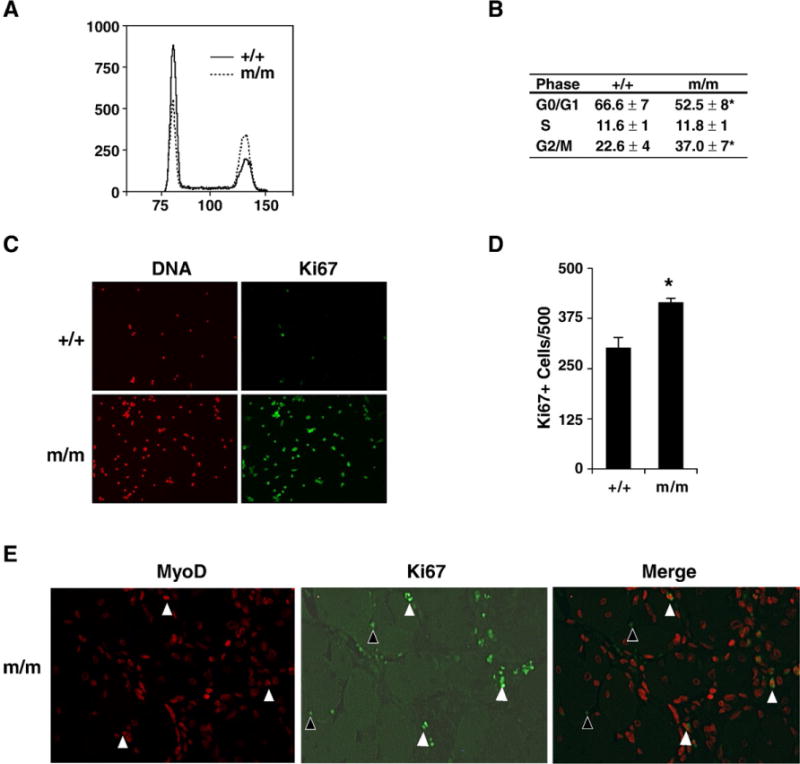
Foxj3m/m MPCs have perturbed cell cycle kinetics. (A) Flow cytometry profile of wild type and Foxj3m/m MPCs reveals that mutant MPCs have decreased number of cells in the G0/G1 stage and increased cells in the G2/M stage of the cell cycle. (B) Table outlines quantitative results of experiments undertaken in panel A representing the average cell cycle kinetics of WT and Foxj3m/m MPCs (n=3 separate experiments; *p<0.003). (C) Ki67 immunostaining of WT and mutant injured skeletal muscle reveals increased proliferation in the absence of Foxj3 following injury. (D) Quantitation of the Ki67 immunostaining reveals approximately a 65% increase in cellular proliferation in the Foxj3m/m injured skeletal muscle (*p<0.05). (E) Immunohistochemical staining of MyoD (red) and Ki67 (green) expression following injury. White arrowheads indicate that a population Ki67 positive cells are also positive for MyoD expression. A sub-population of cells are also positive for Ki67 (black arrowheads) but not myogenic (MyoD negative) in regenerating skeletal muscle.
