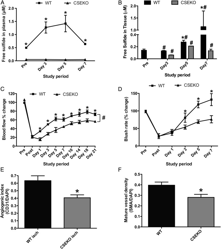
Figure 1.
Blood perfusion and blood vessel density is impaired in CSE KO mice under ischaemia. (A and B) Comparison of WT and CSE KO mice plasma- and tissue-free sulfide levels, respectively, at different time points following hind limb ischaemia induction. (C) Per cent change in ischaemic limb blood flow between WT and CSE KO mice as measured by laser Doppler flowmetry. (D) Per cent change in ischaemic limb indocyanine green (ICG) blush rate between WT and CSE KO mice. (E) Immunohistochemical staining for vascular angiogenic index (CD31/DAPI) between WT and CSE KO ischaemic muscle. (F) Immunohistochemical staining for arterial vessels (SMA/DAPI) between WT and CSE KO mice. n = 7 per cohort, *P < 0.05, #P < 0.05 CSEKO vs. WT.

