Figure 1.
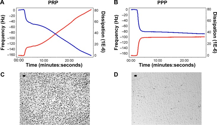
Quartz crystal microbalance with dissipation. Perfusion of PRP leads to platelet microaggregation.
Notes: (A, B) Representative traces for frequency f and energy dissipation D (from the third overtone) recorded by the device. Frequency is represented as a blue line and its values shown in the left axis. Energy dissipation is represented as a red line and its values are shown in the right axis. Perfusion of fibrinogen-coated polystyrene-coated quartz crystals with PRP results in platelet deposition as shown by changes in D (80) and f (−160). In contrast, perfusion with PPP leads to lower changes in D (35) and f (−80), reflecting protein deposition. (C, D) Representative micrographs of the surface of fibrinogen-coated polystyrene-coated quartz crystals as viewed by optical microscopy showing accumulation of platelet microaggregates following perfusion with PRP, but not with PPP. Scale bar 20 μm.
Abbreviations: PRP, platelet-rich plasma; PPP, platelet-poor plasma.

