Figure 7.
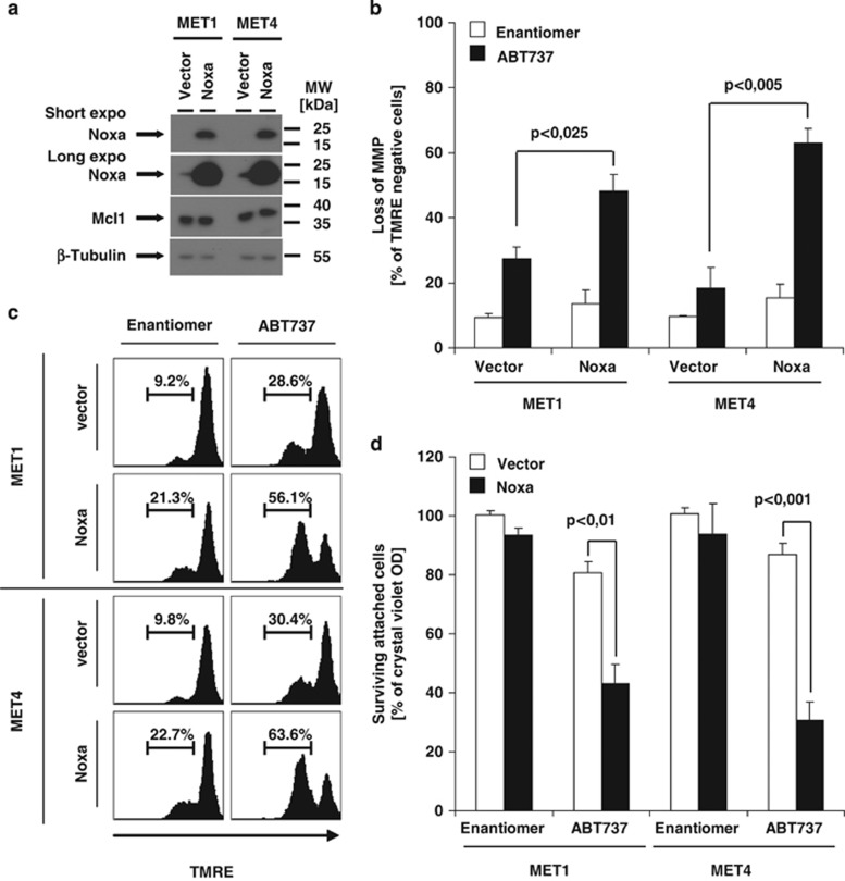
Overexpression of Noxa sensitizes MET1 and MET4 cells to ABT737-induced cell death. (a) Overexpression of Noxa in MET1 and MET4 cells was performed as described in the Materials and Methods section. The successful overexpression of Noxa (a) was determined by western blot analysis using Noxa-specific antibodies; β-tubulin serves as control for equal loading. (b) For analysis of ABT737-induced cell death, cells characterized in (a) were stimulated with diluents (DMSO), Enantiomer (10 μM) or ABT737 (10 μM) for 18–24 h. Surviving attached cells were quantified with crystal violet assay. Summary of three to five independent experiments is shown and S.E.M. is depicted. (c and d) For determination of loss of MMP, the respective genetically manipulated cells were stimulated for 8 h with Enantiomer (10 μM) or ABT737 (10 μM); loss of MMP was visualized by TMRE staining and FACS analysis. One representative experiment (c) or the quantitative summary of three independent experiments together with S.E.M. is shown (a and d)

