
Figure 6. TCDD reduces cell proliferation in adult hippocampus without inducing cell death.
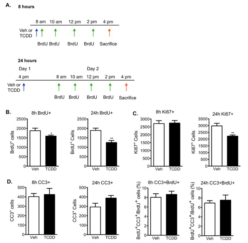
(A)Paradigm used to investigate neural cell proliferation in the dentate gyrus 8h and 24h following TCDD exposure. (B) Quantification of BrdU+ cells in the SGZ demonstrated a reduction in S-phase entry 8h after TCDD exposure compared to vehicle-treated animals, which became more pronounced at 24h. (C) Quantification of Ki67-positive cells revealed a significant reduction in newborn cells in all phases of the cell cycle following 24h exposure to TCDD, but not at 8h. (D) Quantification of CC3-single and CC3- and BrdU-double positive cells relative to total number of BrdU-positive cells in the SGZ demonstrated that TCDD does not induce apoptosis at either timepoint. All results are expressed as means ± S.E.M. (N=10 mice/group); Student's t-test (* p<0.05, ** p <0.01).