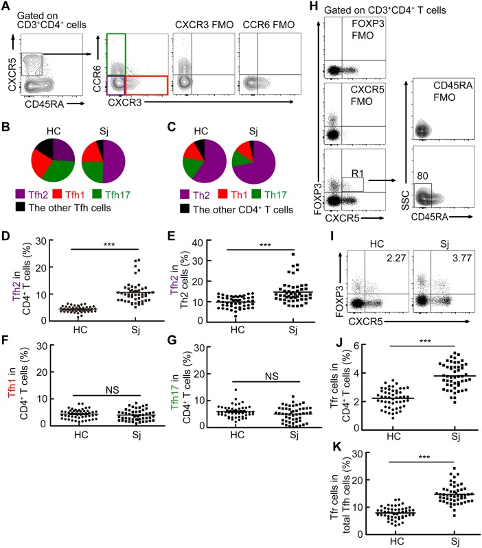
Fig 2. Tfh2 cells and Tfr cells, but not Tfh1 or Tfh17 cells, were significantly increased in patients with schistosomiasis japonica.
(A) Gating schemes for analysis of the percentages of activated Tfh1, Tfh2 and Tfh17 cells. PBMCs were stained with CD3, CD4, CD45RA, CXCR5, CXCR3, and CCR6 antibodies. Gating strategy defining Tfh1 (CXCR3+CCR6- Tfh cells), Tfh2 (CXCR3-CCR6- Tfh cells), and Tfh17 cells (CXCR3-CCR6+ Tfh cells); (B) Shown is the distribution of Tfh1 (red), Tfh2 (purple), Tfh17 (green), and the other subsets (black) within total Tfh cells in healthy controls and patients with schistosomiasis japonica. Areas represent the means of percentages of Tfh1, Tfh2, Tfh17 or the other subsets with CR45RA-CXCR5+ cells. Gated to CR45RA-CXCR5+CD4+ T cells; (C) Shown is the distribution of total Th1 (CXCR3+CCR6- CD4+ T cells, red), Th2 (CXCR3-CCR6- CD4+ T cells, purple), Th17 (CXCR3-CCR6+ CD4+ T cells, green), and the other CD4+ T subsets (black) within total CD4+ T cells in patients with schistosomiasis japonica. Areas represent the means of percentages of Th1, Th2, Th17 or the other CD4+ T subsets with CD4+ T cells; (D-E) Frequencies of Tfh2 with CD4+ T cells (D) or total Th2 cells (E) in healthy controls or patients with schistosomiasis japonica; (F-G) Frequencies Tfh1 (F), and Tfh17 (G) cells within CD4+ T cells in healthy controls or patients with schistosomiasis japonica. (H) Gating schemes for analysis of the percentage of Tfr cells. PBMCs were stained with CD3, CD4, CD45RA, CXCR5, and FOXP3 antibodies. Gating strategy defining Tfr cells (R1); (I-K) Flow cytometry data plots and statistics show the Tfr cells within CD4+ T cells (I, J) or total Tfh cells (K) from one representative individual in indicated groups. All flow cytometry results were analyzed and plotted using Fluorescence Minus One controls (FMO). ***P<0.001, NS indicating not significant.

