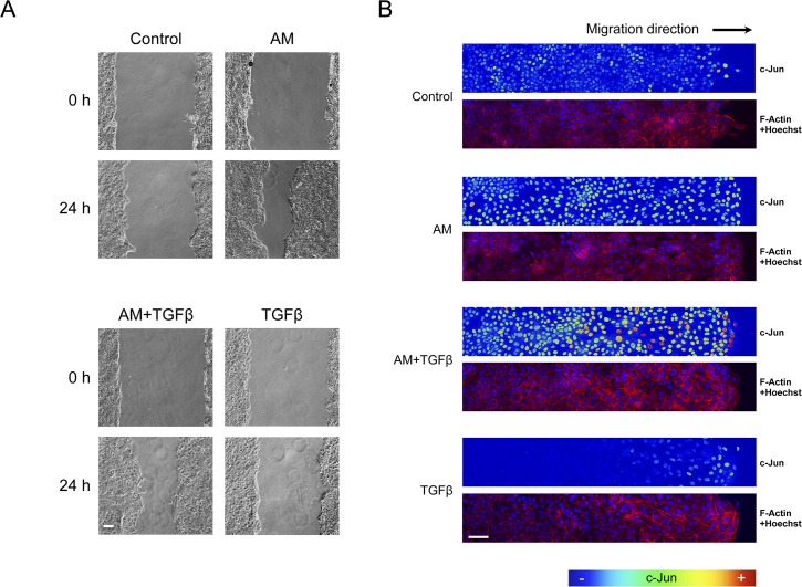
Fig 8. AM induced c-Jun expression is enhanced in the presence of TGFß.
Wound healing scratch assay was performed in HaCaT cells in the presence of AM, TGFß or a combination of both. (A), cells forming a confluent epithelium were wounded and immediately treated as indicated for 24 h. Representative pictures were taken at the beginning of the treatment and 24 h later. (B), wound healing scratch assay was treated with AM, EGF or combinations of AM with different inhibitors. Cells were wounded and treated for 24 h, afterwards cells were fixed and immunostained for c-Jun. Images of c-Jun fluorescence were converted into pseudo-colour to show the intensity of c-Jun staining. Colour rainbow scale represents fluorescence intensity for c-Jun. Co-staining with phalloidin and Hoechst-33258 was used to show the cell structure and nuclei, respectively. Images were taken by confocal microscopy using a Zeiss 510 LSM confocal microscope. This experiment was repeated at least three times. A representative result is shown. Scale Bars 100μm.

