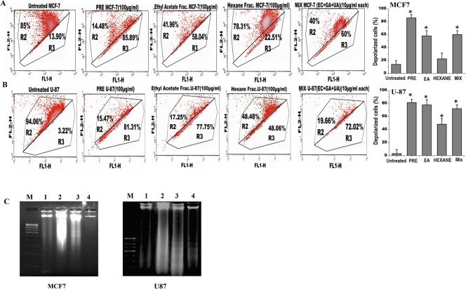
Fig 4. Analysis of apoptosis in (A) MCF7 and (B) U87 cells by measuring the mitochondrial membrane potential after JC1 staining in untreated and treated with PRE, EA, Hex fractions and mixture of EC+GA+UA of EA fraction of P. fulgens root.
The upper part indicates the percentage of cells show polarization of mitochondrial membrane and the lower part shows the percentage of cells having mitochondrial membrane depolarization. All these experiments were repeated twice. Right panel shows percentage of depolarized cells after each treatment. *p<0.05, Students’ t-test as compared with untreated control. (C) Effect of PRE, Hexane and ethyl acetate soluble fraction treatment on the DNA degradation and fragmentation in MCF7 and U87 cells. DNA samples are labeled as: (M) molecular weight marker, (1) untreated cells, (2) cells treated with hexane-fraction, (3) cells treated with PRE and (4) cells treated with ethyl acetate fraction. Results are representative of two independent experiments. The concentration was used 100 μg/ml and the treatment was given for 24 h.

