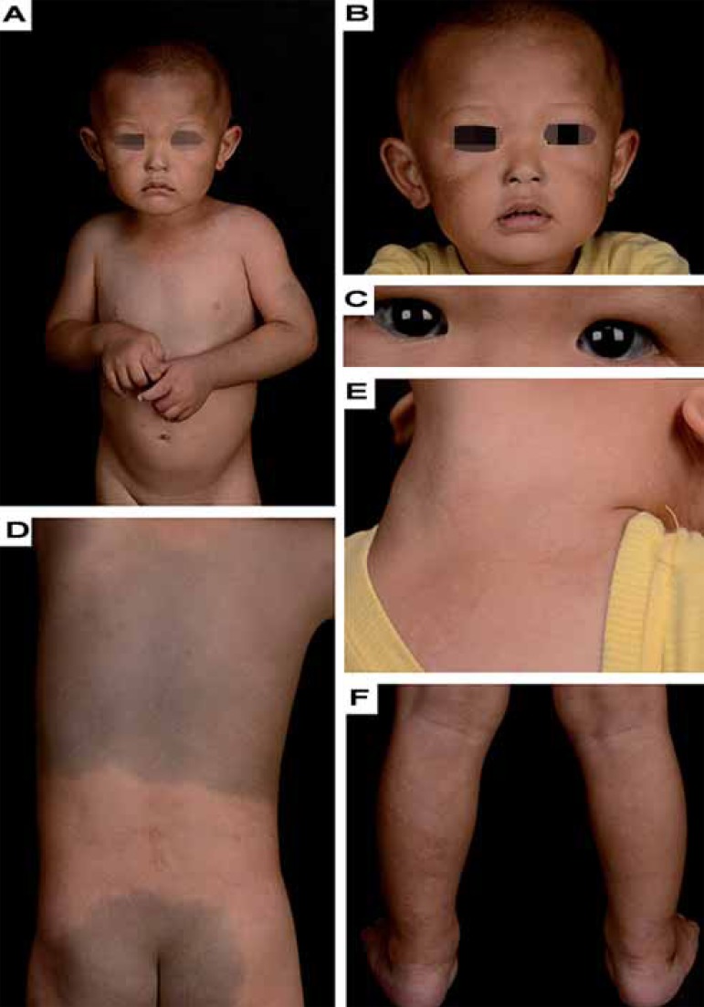
FIGURE 1.
(A and B) Bilateral nevus of Ota on the face. (C) Blue spots on the cornea and conjunctiva. (D) Demarcated greyish-blue hyperpigmentation over the back and buttocks. (E) Erythematous patches on the regio cervicalis anterior. (F) Localized reticulated erythema and bluish-grey macules consistent with irregular pale macules and patches over the left leg calf; note that pale macules and patches might be more obvious on the rear side of the right ankle

