FIGURE 3.

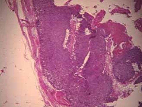
Lesion lined by a basaloid epithelium at the periphery, filled with eosinophilic cornified material and shadow cells (HE 200x)

Lesion lined by a basaloid epithelium at the periphery, filled with eosinophilic cornified material and shadow cells (HE 200x)