
Fig 2. Deletion of Dclk1 exacerbates colonic barrier dysfunction following DSS treatment.
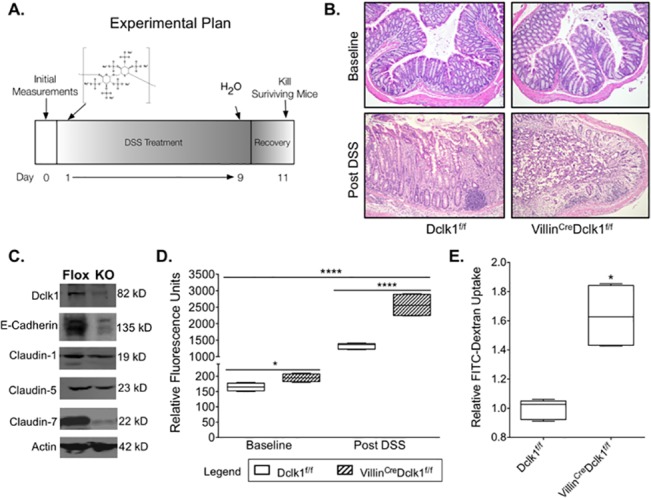
A. Experimental plan; B. H&E staining demonstrated normal histology in VillinCre;Dclk1f/f and Dclk1f/f mice at baseline and confirmed a significant inflammatory response following DSS treatment (200x magnification) C. Western blot analysis of tight junction and adherens junction proteins demonstrated a decrease in Claudin-1, Claudin-7, and E-cadherin in the VillinCre;Dclk1f/f mice after DSS treatment; D. FITC-Dextran levels in the serum of each mouse were determined 4 h after gavage in baseline and post DSS treatment (n = 5 for post DSS groups, *p<0.02, **** p<0.0001 VillinCre;Dclk1f/f vs Dclk1f/f); E. Fold change of FITC-Dextran levels in the VillinCre;Dclk1f/f mice post DSS treatment.