
Fig. 4.
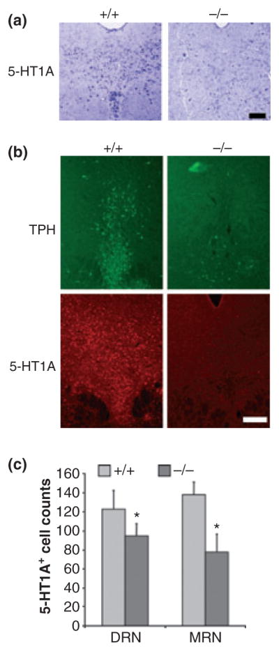
Reduced 5-HT1A mRNA and protein expression in Pet-1−/− raphe nuclei. (a) In situ hybridization showing 5-HT1A mRNA anti-sense probe staining in the dorsal raphe of wild-type (+/+) and Pet-1 knockout (−/−) mice; scale bar = 50 μm. (b) Immunofluorescent labelling for tryptophan hydroxylase (TPH) or 5-HT1A receptor proteins in sections of DRN from the same wild-type or knockout mice. Representative images are shown at 10× magnification with a scale bar = 100 μm; n = 3 mice/group. (c) Quantification of 5-HT1A-immu-nopositive cells. Cell counts are presented from the dorsal (DR) and median raphe (MRN) of wild-type (+/+) and Pet-1 knockout (−/−) mice; n = 3 mice/group. Statistical significance was measured using two-tailed unpaired t-test, where *p < 0.05.