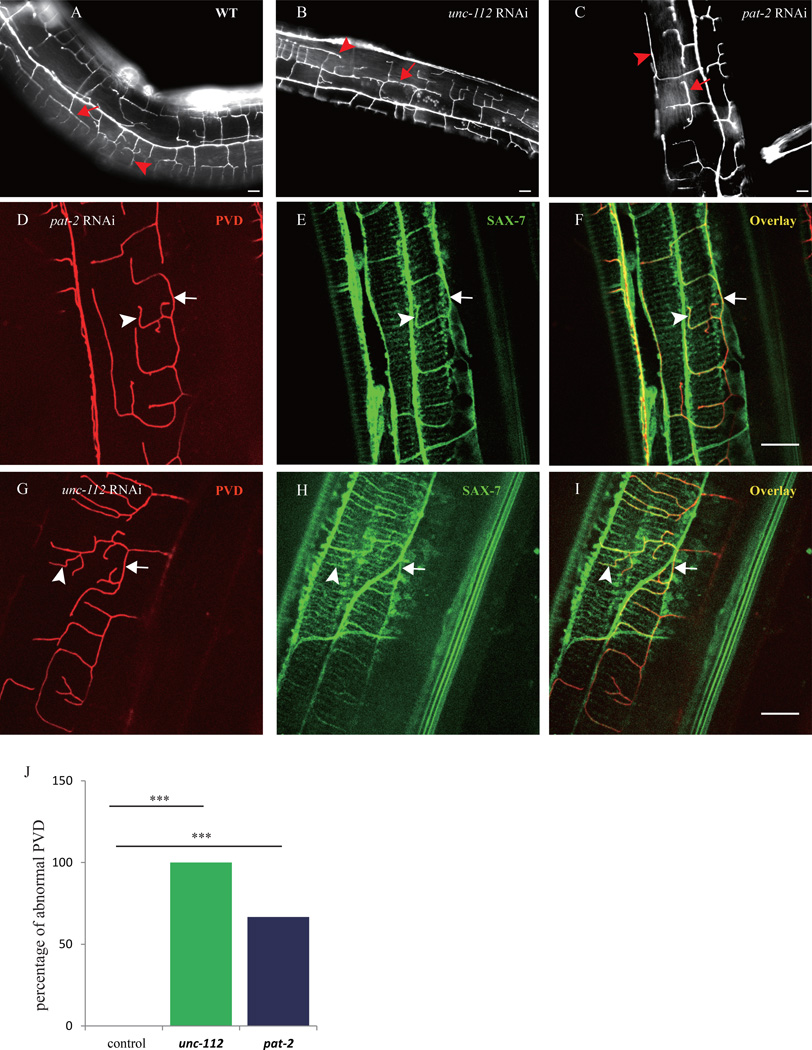
Fig. 4. Sarcomere components are important for the SAX-7 pattern and PVD branch morphology.
A–C) Florescent images of PVD in young adult wild type and unc-112 (RNAi), pat-2 (RNAi) animals. D–I) Confocal images of PVD and Pdpy-7::SAX-7::YFP of the unc-112 (RNAi), pat-2 (RNAi) animals. Arrows indicate the 3° branches and the SAX-7 lateral stripes. The arrowheads indicate the 4° branches and the co-localized SAX-7 stripes. J) Quantification of the RNAi penetrance. n>30 for each genotype. ***p < 0.001 by x2 test. Scale bar is 10 µm.

