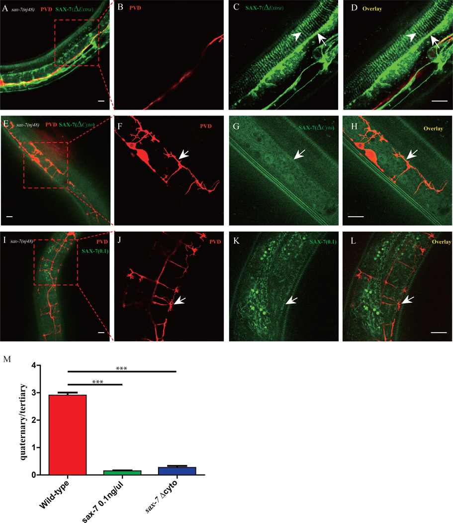
Fig. 5. Structure-function analysis of SAX-7.
A–D) Confocal images of the Pdyp-7::SAX-7(ΔExtra)::YFP transgene in sax-7(nj48) mutant. n>30. E–H) Confocal images of the Pdyp-7::SAX-7(ΔCyto)::YFP transgene in sax-7(nj48) mutant. n>30. I–L) Confocal images of the Pdyp-7::SAX-7::YFP(0.1ng/µl) transgene in sax-7(nj48) mutant. n>30. M) Quantification of rescue activity of SAX-7 truncation constructs in A) and I). Error bars, SEM. ***p < 0.001 by Student’s t test, n>10 for each genotype. Scale bar is 10 µm.

