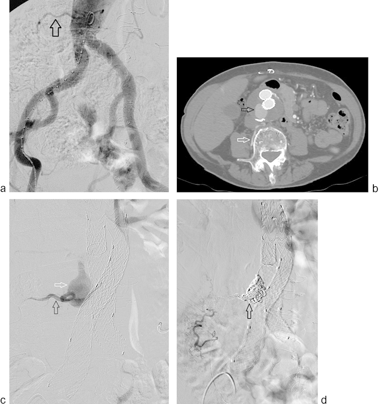
Fig. 1.

Pre-EVAR planning angiogram (a) demonstrates a patent right lumbar artery (arrow). Postoperative year-4 surveillance CT angiogram (b) shows a patent right lumbar artery (white arrow) and type 2 endoleak (black arrow) with significant growth of the aneurysm sac. Selective right lumbar angiogram (black arrow) (c) shows filling of the aneurysm sac (white arrow). Completion angiogram (d) following successful coil embolization (arrow) of the aneurysm sac and right lumbar artery.
