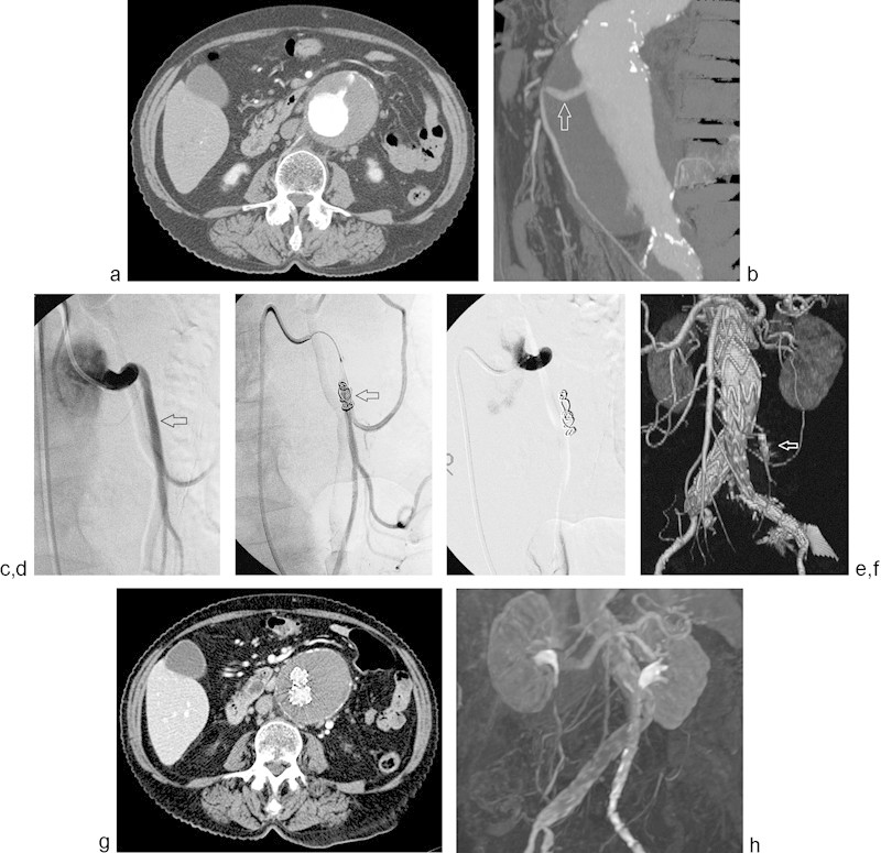
Fig. 2.

Axial CT angiogram (a) demonstrates a large infrarenal AAA with a widely patent IMA. 3D CT angiogram reconstruction (b) re-demonstrates a patent IMA (arrow). Pre-EVAR angiogram (c) demonstrates a patent IMA (arrow). Platinum coils were deployed with intermittent angiography (arrow) (d) until complete IMA stasis was achieved (e). CT angiogram 3D reconstruction (f) obtained 1 month post-EVAR demonstrates occlusion of the IMA (arrow) and absence of endoleak. Surveillance CT (g) and MR (h) angiogram, obtained at 2 and 3 years postoperatively, respectively, again demonstrate freedom from type 2 endoleak or sac enlargement.
