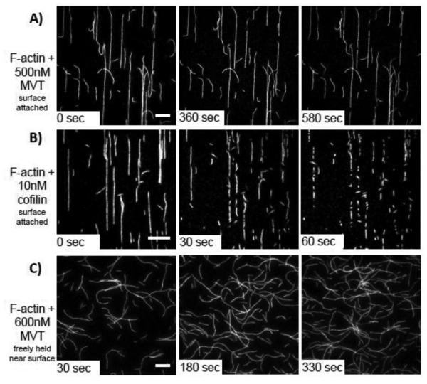
Figure 2. MVT does not actively sever actin filaments.
A-B) 1μM Mg-ADP-F-actin (20% Cy3b-labeled, 0.5% Biotin-Peg2-labeled) was attached to biotin-PEG on functionalized coverslips through streptavidin cross-links. Once an initial image was acquired (0 sec), the indicated proteins were added and movies were acquired. A) Very few breaks were observed when MVT was added (see also in Supplementary Movie 1). B) Cofilin rapidly severs filaments under the same conditions (see also in Supplementary Movie 2). Scale bars = 10 μm. Cofilin images are shown at a higher magnification to better visualize the large number of severing events. C) F-actin (20% Cy3b-labeled) was diluted to 25 nM in the presence of 0.6 μM MVT (binding density = 0.5) and immediately added to chambers treated with 1% Pluronic F-127 and 0.5 mg/ml casein. In this experiment, the filaments are not covalently attached to the surface. No cuts were detected. Scale bar = 20 μm.

