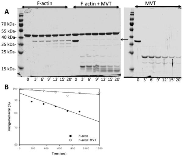
Figure 6. Limited proteolysis of MVT decorated F-actin by subtilisin.
A) 10 μM F-actin, 10 μM MVT, and 10 μM F-actin+MVT were all digested with the same concentration of subtilisin, based on enzyme to protein mass ratio of 1:50 (subtilisin:F-actin). Reaction aliquots were taken at the indicated time points and were analyzed by 12% SDS/PAGE gels. B) Digestion of actin monomer was analyzed by densitometry of intact actin monomer band. MVT protects actin’s D-loop from proteolytic cleavage by subtilisin.

