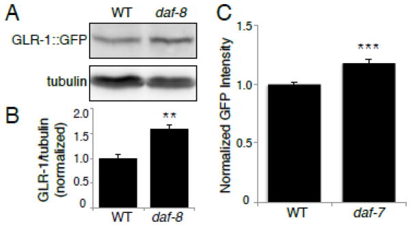
Figure 5. GLR-1::GFP protein levels and promoter activity are increased in DAF-7/TGF-β pathway mutants.

(A) Immunoblot analysis of protein levels in wild type (WT) and daf-8(e1393) worms. The proteins analyzed are GLR-1::GFP (anti-GFP antibody) and tubulin (anti-β-tubulin antibody). (B) Protein levels were quantified using densitometry, shown is the quantification of GLR-1::GFP levels normalized to tubulin, n=6. (C) Maximum GFP fluorescence intensity was measured in the nucleus of PVC neurons of WT and daf-7(e1372) worms expressing Pglr-1::NLS-LacZ-GFP. (WT: 1.00±0.02 SEM, n=78) (daf-7: 1.18±0.02 SEM, n=77). Error bars denote SEM. ** p<0.01, *** p<0.001 (Student’s t-test).
