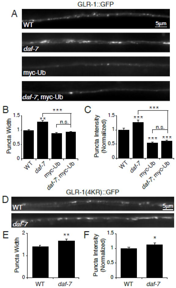
Figure 6. Ubiquitin-dependent degradation of GLR-1 in daf-7 mutants.
(A) Representative images of GLR-1::GFP in the VNC of wild type (WT), daf-7(e1372), worms overexpressing myc-Ub (Pglr-1::myc-Ub), and daf-7(e1372); Pglr-1::myc-Ub. (B–C) Quantification of puncta width (B) and normalized fluorescence intensity (C) of WT n=38, daf-7(e1372) n=28, myc-Ub n=29, and daf-7(e1372); myc-Ub n=38. (D) Representative images of GLR-1(4KR)::GFP in the VNC of WT and daf-7(e1372) worms. (E–F) Quantification of puncta width (E) and normalized fluorescence intensity (F) of WT n=28 and daf-7(e1372) n=21. Error bars denote SEM. * p<0.05, ** p<0.01, *** p<0.001 (Tukey-Kramer test).

