
Figure 1. Pre-operative axial MR imaging of deep brain nuclei after cochlear implant magnet removal.
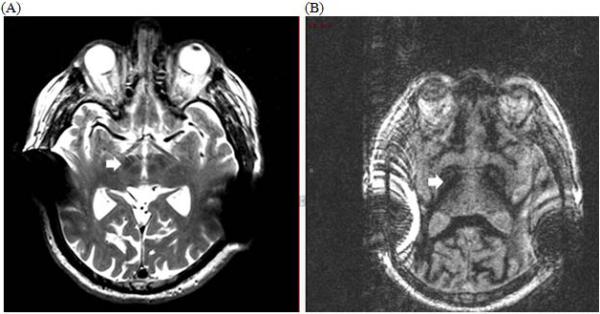
(A) Axial T2-weighted MRI Turbo Spin-Echo sequence depicting subthalamic nuclei (arrow) (B) Axial MRI Fast Gray Matter Acquisition T1 Inversion Recovery sequence depicting globus pallidus internus (arrow), but the image is significantly degraded with artifact from the cochlear implants despite magnet removal.