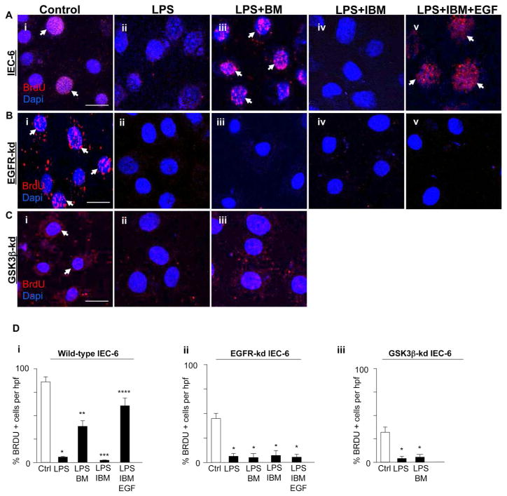
Figure 7. Breast milk restores enterocyte proliferation via EGFR and GSK3β.
A–C: Representative confocal micrographs of either wild-type IEC-6 cells (Ai–v) or EGFR-kd IEC-6 (Bi–v) or GSK3β-kd IEC-6 cells (Ci–iii), treated as indicated, then stained for the proliferation marker bromodeoxyuridine (BrdU, red, arrows) and DAPI (blue). D: Quantification of the BrdU positive cells per high power field (hpf) in wild-type IEC-6 cells (i) or EGFR-kd IEC-6 (ii) or GSK3β-kd IEC-6 cells (iii). *p<0.05 versus control (Ctrl, white bar), **p<0.05 versus LPS, ***p<0.05 versus LPS + breast milk (BM), ****p<0.05 versus LPS + IBM. Results representative of three separate experiments with over 50 high power fields per group. Size bar = 10μm. Data are mean±SEM. Arrows delineate proliferative cells.

