Figure 6.

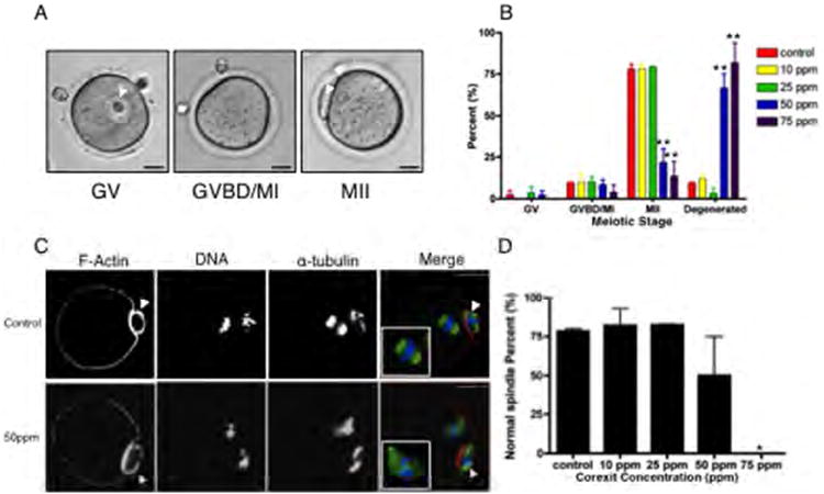
CE exposure affects oocyte meiotic competence and spindle morphology. The ability of oocytes derived from follicles exposed to CE to progress through the stages of meiotic maturation was (A) scored by light microscopy and (B) quantified. Cells were classified as germinal vesicle intact (GV), germinal vesicle breakdown/metaphase of meiosis I (GVBD/MI), or metaphase of meiosis II (MII), and representative images for each stage are shown (A). This experiment was repeated at least twice and a minimum of 25 cells were analyzed in each experimental group. (C) The actin- and microtubule-based cytoskeleton in the resulting MII eggs from control and CE-exposed follicles was examined by immunocytochemistry and confocal microscopy. An optical section encompassing the meiotic spindle is shown. In the merged images, the pseudocolor represents the following: green=α-tubulin, red=F-actin, and blue=DNA. Normal meiotic spindles were characterized by a bipolar structure with chromosomes tightly aligned on the metaphase plate (upper panel, control). Abnormal spindles were characterized by disrupted tubulin and scattered chromosomes (lower panel, 50 ppm CE). (D) The percent of normal spindles observed in the MII eggs derived from follicles exposed to increasing concentrations of CE was quantified. This experiment was repeated a minimum of 2 times, and between 18 and 29 oocytes in each experimental condition were analyzed. Representative images are shown. Arrowheads indicate the polar body. Scale bar=25μm. Statistically significant differences are denoted as follows: *P<0.05.
