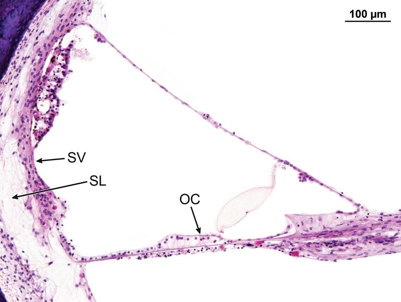
Figure 5.
Organ of Corti in the middle turn (25mm from the basal end of the cochlea) of Case #1, corresponded to boxed area in Fig. 3. The organ of Corti was replaced by a mound of epithelial cells in which there were no obvious hair cells present. There was atrophy of the stria vascularis (SV) and spiral ligament (SL).

