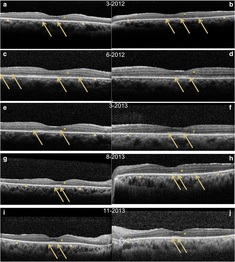
Fig. 2.

Fourier-domain cross-sectional optical coherence tomography (Spectralis) that shows initial disruption in the outer nuclear layer (ONL) and external limiting membrane (ELM) and decreases in reflectivity of the inner segment ellipsoid (ISe) in the a right eye and b left eye on March 2012. Arrows point to changes in the ISe, and arrowheads show tapering of the ONL. c, d All layers recover in the immediate follow-up visit on June 2012. e–j FD-OCT shows loss of the ONL and progressive decrease in reflectivity of the ISe with eventual loss of the layer in the final images. i, j Abnormal thickening of the nerve fiber layer. The ONL shows abnormal hyperreflectivity throughout the inner half on all images (asterisk)
