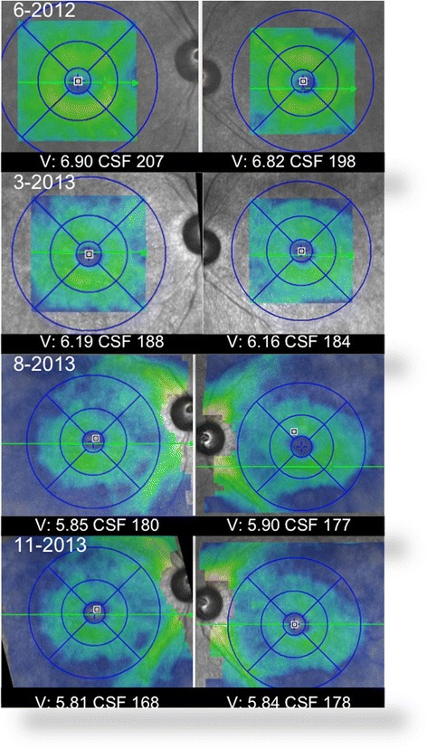
Fig. 3.

The volume map (Spectralis) from each visit shows progressive retinal atrophy. Right eye volume (V) on the listed dates is 6.90, 6.19, 5.85, and 5.81 mm3, and the left eye volume is 6.82, 6.16, 5.90, and 5.84 mm3. Central subfield thickness (CSF) is recorded on each image
