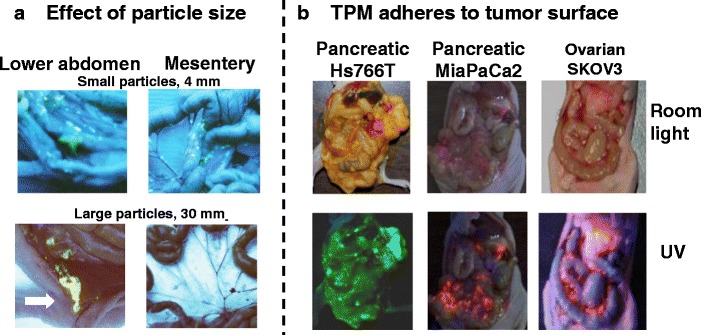
Fig. 3.

Intraperitoneal distribution of PLGA microparticles. a Effect of particle size. Tumor-free mice were given IP injections of acridine orange-labeled PLGA microparticles with average diameters of 4 or 30 μm. Acridine orange appears yellow under UV light. The smaller particles were dispersed throughout the cavity, and on mesenteric membrane and omentum that are common sites of local metastases of ovarian tumors. The larger particles were localized in lower abdomen near the injection site (indicated by an arrow) and were absent on mesenteric membrane and omentum. b TPM (4–6 μm) adheres to tumor surface. Mice were implanted with IP human xenograft tumors (pancreatic Hs766T, pancreatic MiaPaCa2, or ovarian SKOV3). After tumors were established (day 21, 28, and 42, respectively), mice were given an IP dose of FITC- or rhodamine-labeled blank TPM. Three days later, the animal was anesthetized and the abdominal cavity exposed. Green or red color under UV light indicated localization of FITC and rhodamine, respectively. Reprinted from (6) with permission
